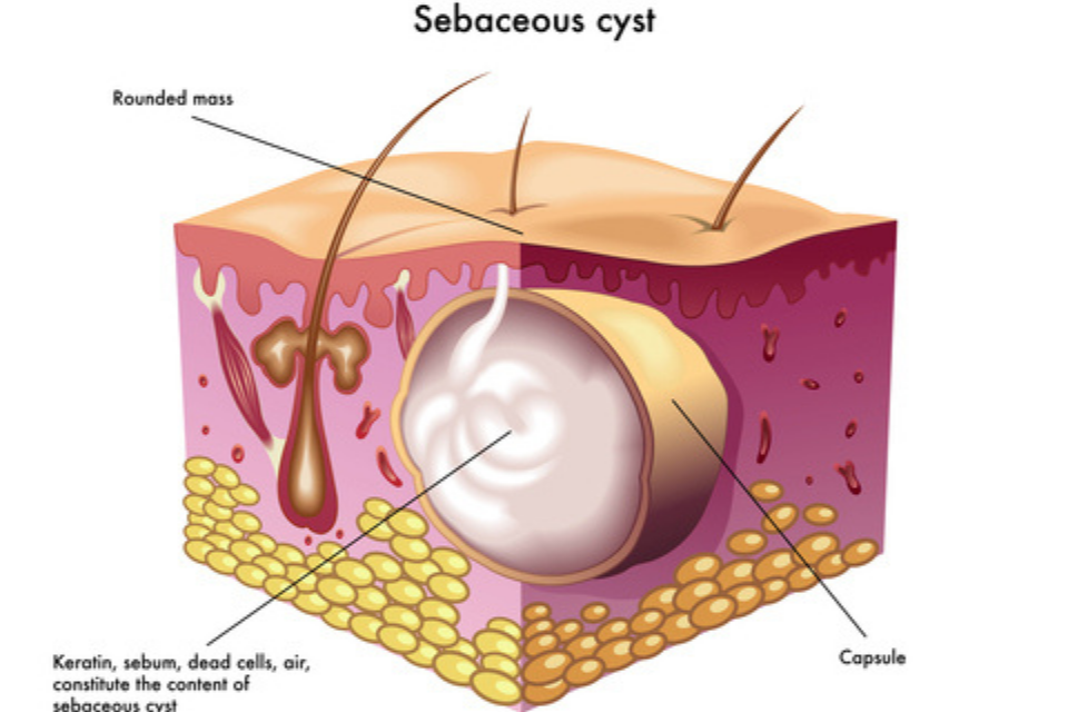
Sebaceous cysts, by definition, are not malignant and have zero malignant potential. However, they can become infected if bacteria gets trapped within this sack of dead skin.
Normally, sebaceous cysts are asymptomatic. If the cyst partially ruptures, and dead skin ends up in the surrounding tissue, a foreign body reaction can result. This looks like a red, lump of skin and can be very tender. As mentioned earlier, if bacteria gets into this closed space or sac, an abscess can result that may be very tender and often requires drainage.
Removing sebaceous cysts is quite simple. We numb up the skin in the area, nick the top of the skin with a small blade and remove the entire cyst sac outside of the skin.
“Sebaceous cysts” are one of the most common skin conditions that we treat in dermatology. Patients notice lumps underneath their skin that may be smaller than a centimeter, but occasionally can be much larger. They feel semi-firm and are usually skin colored. Classically, we are able to see a small ‘dot’ somewhere on the lump, which is a clue that it is a “sebaceous cyst” rather than another type of lump.
The term “sebaceous cyst” is actually an inaccurate name that has kind of just stuck over time. This name suggests that the cyst is made up of oily secretions from the sebaceous glands, when in actual fact, the cyst actually contains layers of ‘keratin’, a protein that is part of our skin and hair follicles. The more appropriate medical name for them is therefore ‘epidermoid cysts’.
Epidermoid cysts occur when there is disruption in the upper layer of a hair follicle. In normal circumstances, a hair follicle opens up onto the surface of the skin without interruption. In an epidermoid cyst, a disruption to this hair follicle causes a blockage at the opening of the follicle, and therefore a build up of the normal skin/hair keratin to form behind it. This means that epidermoid cysts do not form on areas without hair follicles e.g. palms of the hands and soles of the feet.

They are not dangerous in the majority of cases. Most of the time, these blockages of the hair follicles don’t even cause symptoms and patients simply notice a lump. Occasionally, when these cysts rupture underneath the skin, the keratin inside them leaks into the surrounding tissue and causes inflammation. In this case, they can become painful/red.
Also, bacteria can occasionally get inside the cyst and cause an infection. This would also result in some pain, redness and inflammation being visible on the skin.
The lumps caused by true epidermoid cysts are not a type of skin cancer, and do not become cancer other than in exceptionally rare circumstances.
There are multiple options available to treat epidermoid cysts, but cutting the entire cyst out using a small surgery has the highest rates of curing the problem without it coming back. This involves numbing the area using a numbing injection, followed by a small cut with a blade. We then remove the entire cyst with the lining to make sure that the lump doesn’t simply re-form, and then close the skin using stitches.
When the cyst is very inflamed or we are concerned for infection, we often treat patients with a course of antibiotic tablets to help bring down the redness and inflammation before doing a full surgery to cut out the cyst. This is because the surgery is easier to do and results in better healing often when the cyst is not red and inflamed.
Other treatment options include injecting the cyst with a steroid solution, and also making a small cut in the skin and squeezing out some of the cyst contents. Both of these can help to reduce inflammation and the size of the cyst, but because they don’t remove the wall of the cyst (where the proteins come from), the cyst can often form again within months to years. They are a good option for people who don’t want surgery with stitches, but just a short to medium term fix for the problem.
Although epidermoid cysts are a very common cause of lumps in the skin, there are other conditions that can present in a very similar way. Under the skin lumps can occasionally be caused by other types of hair follicle cysts, or also be collections of fat cells known as ‘lipomas’. The best way to figure out which type of cyst you may have is by being evaluated by a dermatologist. We can identify where your cyst is (different types of cyst are more common in different locations), what it looks like under magnification (does it have a hair follicle visible which can be a clue to an epidermoid cyst), is it moveable (this can be more typical of a lipoma), and many others aspects. Also, if we remove the cyst with a surgery, we send this to the lab to get a definite diagnosis and provide you peace of mind.